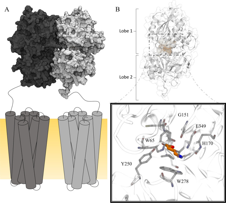
Figure 1.
Schematic illustration of the GABAB-R. The heterodimeric GABAB-R is comprised of GABAB1a/b (gray) and GABAB2 (black) (A). The 7TM domains are located in the membrane (yellow) (A). The orthosteric binding site is located in the extracellular VFT of GABAB1a/b (B). Ligand binding is facilitated by interactions with key residues such as Tyr250 and Trp278 located in Lobe 2, Gly151, His170, and Glu349 located in Lobe 1 (black box, PDB ID: 4MS3 in complex with GABA).

