Figure 6.

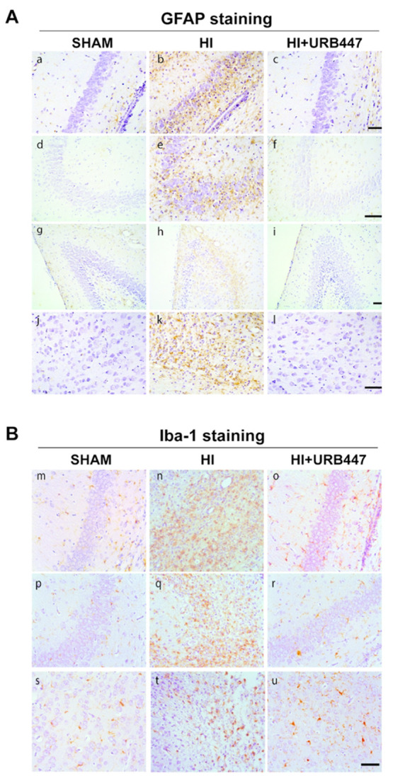
Modulation of glial fibrillary acidic protein (GFAP) and ionized calcium binding adaptor molecule-1 (Iba-1) staining by neonatal hypoxia-ischemia (HI) and treatment with URB447. (A) Representative light microphotographs illustrating GFAP immunostaining in brain sections from CA1 (a–c) and CA2/CA3 (d–f) hippocampal regions, dentate gyrus (g–i) and cortex (j–l). Samples were obtained from 14-day-old rats subjected to sham operation (SHAM; a, d, g, j) or to HI on P7 and treated with either vehicle (HI; b, e, h, k) or URB447 (HI+URB447; c, f, i, l). (B) Representative light microphotographs illustrating Iba-1 immunostaining in brain sections from CA1 (m–o) and CA2/CA3 (p–r) hippocampal regions and cortex (s–u). Samples were obtained from 14-day-old rats subjected to sham operation (SHAM; m, n, o) or to HI on P7 and treated with either vehicle (HI; n, q, t) or URB447 (HI+URB447; o, r, u). Bars, 100 μm.
