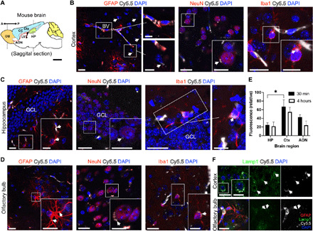
Fig. 2. Neural cell uptake of St-Cl-Pr-Cy5.5-ANG after passage through the BBB.

(A) Schematic of an adult mouse brain representing the two antero-posterior coronal levels studied for the presence of St-Cl-Pr-Cy5.5-ANG. AON, anterior olfactory nucleus; Ctx, cerebral cortex; CB, cerebellum; CC, corpus callosum; HP, hippocampus; LV, lateral ventricle; OB, olfactory bulb. (B) The presence of St-Cl-Pr-Cy5.5-ANG in the cerebral cortex, showing abundant accumulation in the blood vessel (BV) walls and surrounding GFAP+ astrocytes (left), and in punctate regions (arrows) in the cytoplasm of NeuN+ neurons (middle) and Iba1+ microglia (right). (C and D) Distribution of St-Cl-Pr-Cy5.5-ANG (arrows) in GFAP+ astrocytes (left), NeuN+ neurons (middle), and Iba1+ microglia (right) in the granule cell layer (GCL) of the dentate gyrus of the hippocampus (C) and the olfactory bulb (D). (E) Quantification of Cy5.5 fluorescent intensity between different brain regions at short (30 min) and long (4 hours) time points after intravenous administration of St-Cl-Pr-Cy5.5-ANG. All data shown are relative fluorescence intensity values corrected for background autofluorescence of the same brain regions in vehicle-injected mice (means ± SEM; n = 4 mice per time point). Two-way ANOVA followed by Šidák’s post hoc test, *P < 0.05. (F) St-Cl-Pr-Cy5.5-ANG (arrows) localizes to lysosomes (Lamp1, green) in cells of the cerebral cortex (top) and olfactory bulb (bottom). Scale bars, 20 μm in panoramic images and 10 μm in insets.
