
Fig. 4. Analysis of St-Cl-Pr-BDMC capability to inhibit Aβ-induced neurotoxicity and to induce a neurotrophic effect in hippocampal organotypic cultures.
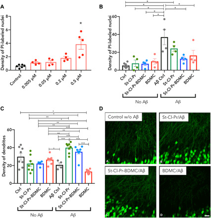
(A) Changes in density (nuclei/1000 μm2) of PI-stained nuclei in the pyramidal layer of the CA1 region of hippocampal organotypic cultures comparing control cultures treated with vehicle and cultures treated with different concentrations of St-Cl-Pr-BDMC (0.005, 0.05, 0.2, and 0.5 μM drug-equivalents). Asterisks indicate statistically significant differences after ANOVA analyses followed Bonferroni’s post hoc tests. n > 3, means ± SEM (20). (B) Changes in the density (nuclei/1000 μm2) of PI-stained nuclei in the pyramidal layer of the CA1 region of hippocampal organotypic cultures when comparing control cultures treated with vehicle and cultures treated with the polymer St-Cl-Pr, St-Cl-Pr-BDMC, or free BDMC (0.05 μM). The distinct groups were subsequently treated with vehicle (no Aβ) or Aβ1–42 peptide (Aβ). Asterisks indicate statistically significant differences after ANOVA analyses followed Bonferroni’s post hoc tests. n > 3, means ± SEM (20). (C) Graph representing the changes in dendrite density of CA1 pyramidal neurons measured in the stratum radiatum (optical density, arbitrary units) of hippocampal organotypic cultures when compared with control cultures treated with St-Cl-Pr, St-Cl-Pr-BDMC, or free BDMC (0.05 μM). The distinct groups were subsequently treated with vehicle (no Aβ) or Aβ1–42 peptide (Aβ). Asterisks in bars indicate statistically significant differences between groups after ANOVA analyses followed by Tukey or Games-Howell post hoc tests. (D) Confocal microscopic analysis of dendrite density of pyramidal neurons in the stratum radiatum of CA1 of hippocampal organotypic cultures. All microphotograph images are derived from single confocal planes. Scale bar, 25 μm.