
Fig. 7. TRPC5 channels are located in the odontoblast layer.
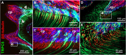
(A) TRPC5 reporter mouse molar tooth whole mount with densely packed TRPC5+ odontoblasts at the pulp-dentin boundary. (B and C) In tight association with sensory nerves. Green, TRPC5; red, βIII-tubulin; circle indicates area shown in (D) and (E) from a subsequent section. cp, coronal pulp; rp, radicular pulp. (E) Oblique section through the predentinal radicular pulp to visualize the tight association of TRPC5+ odontoblast processes with their sensory nerves.