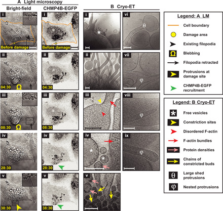
Fig. 6. Live-cell light microscopy and cryo-ET of damage sites in Vps4B knockdown cells.

(A) Light microscopy images of Vps4B knockdown HeLa cells grown on glass (i) before and (ii to v) at various time points after damage—(left) bright-field and (right) CHMP4B-EGFP imaging. EGFP fluorescence is shown using inverted grayscale. The damage area is 3 μm in diameter. (B) Cryo-ET of damage sites of Vps4B knockdown cells showing actin-filled plasma membrane protrusions, pearling/budding profiles, shed vesicles, protein densities observed at certain sites of high membrane curvature in budding profiles and shed vesicles, chains of budding profiles, shed membrane protrusions devoid of F-actin, and nested protrusions. Samples sizes for quantifications: 11 tomograms for Vps4B knockdown (versus 10 tomograms for wild type). Scale bars, 10 μm (A) and 200 nm (B).
