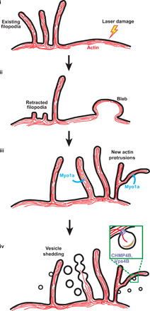
Fig. 7. Model for damage-induced plasma membrane shedding.

(i and ii) F-actin and membrane are redirected to the site of plasma membrane damage from other regions of the cell (possibly from existing filopodia as well) during the formation of membrane blebs at this site. (iii) Membrane blebs are withdrawn into the cell and reorganized into membrane protrusions using F-actin. There is an overall enrichment of protrusions at sites of damage. (iv) F-actin depolymerization could cause membrane deformation and pearling. Proteins that sense or induce high negative membrane curvature could provide additional force necessary for membrane deformation (especially for smaller vesicles). Myo1a is involved in building protrusions and/or shedding dynamics, while Vps4B is involved in membrane scission during shedding of smaller vesicles. Overall, cells likely form new protrusions in response to damage and shed membrane vesicles from these protrusions, both processes occurring predominantly at the site of damage.
