. 2021 Mar 2;299(1):36–48. doi: 10.1148/radiol.2021201948

Figure 1:

Diagram of imaging protocol for contrast-enhanced mammography. Two minutes before image acquisition, iodine-based contrast material is injected. Next, at minimum, both breasts are imaged in craniocaudal and mediolateral oblique views. In each step, compression is applied (red arrow), followed by rapid acquisition of low- and high-energy images. These images are processed to generate low-energy and recombined images. After each exposure, compression is released (green arrow). Images are considered to be of diagnostic value if they are acquired within 10 minutes after contrast material administration. mins. = minutes.

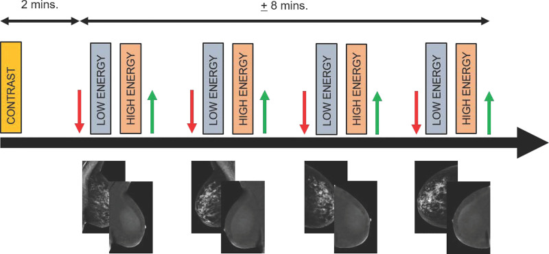
Diagram of imaging protocol for contrast-enhanced mammography. Two minutes before image acquisition, iodine-based contrast material is injected. Next, at minimum, both breasts are imaged in craniocaudal and mediolateral oblique views. In each step, compression is applied (red arrow), followed by rapid acquisition of low- and high-energy images. These images are processed to generate low-energy and recombined images. After each exposure, compression is released (green arrow). Images are considered to be of diagnostic value if they are acquired within 10 minutes after contrast material administration. mins. = minutes.