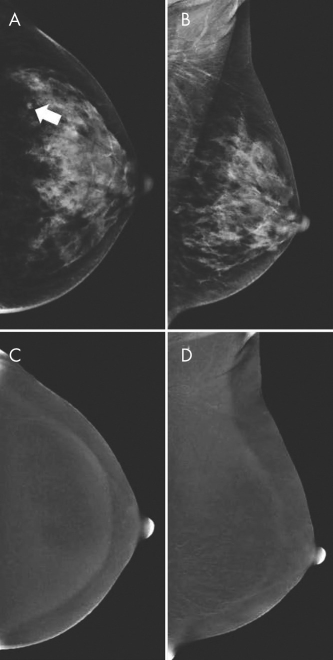
Figure 2:

A, B, Contrast-enhanced mammographic (CEM) images in 52-year-old woman recalled from screening for new ill-defined mass (arrow in A) visible on, A, low-energy craniocaudal view but not visible on, B, low-energy mediolateral oblique view. C, D, Subsequent evaluation of recombined CEM images revealed no suspicious enhancement. Targeted US showed no lesions, but because of small lesion size, patient was categorized as having Breast Imaging Reporting and Data System category 3 lesion (follow-up after 6 months showed no breast cancer). Two subsequent rounds of screening (up to 4 years after primary evaluation) revealed no breast cancer.
