. 2021 Mar 2;299(1):36–48. doi: 10.1148/radiol.2021201948

Figure 3:

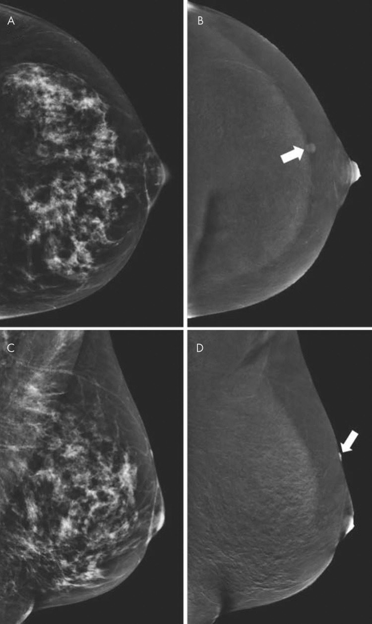
Contrast-enhanced mammographic images of enhancing benign lesion in 50-year-old woman. A, Low-energy craniocaudal image. B, Contrast-enhanced recombined craniocaudal image. C, Low-energy mediolateral oblique image. D, Contrast-enhanced recombined mediolateral oblique image. Contrast-enhanced craniocaudal view of left breast demonstrates a small, well-defined enhancing mass (arrow in B). Mediolateral oblique view of same breast demonstrates that lesion is located on skin (arrow in D). Visual inspection revealed skin hemangioma.

Contrast-enhanced mammographic images of enhancing benign lesion in 50-year-old woman. A, Low-energy craniocaudal image. B, Contrast-enhanced recombined craniocaudal image. C, Low-energy mediolateral oblique image. D, Contrast-enhanced recombined mediolateral oblique image. Contrast-enhanced craniocaudal view of left breast demonstrates a small, well-defined enhancing mass (arrow in B). Mediolateral oblique view of same breast demonstrates that lesion is located on skin (arrow in D). Visual inspection revealed skin hemangioma.