. 2021 Mar 2;299(1):36–48. doi: 10.1148/radiol.2021201948

Figure 5:

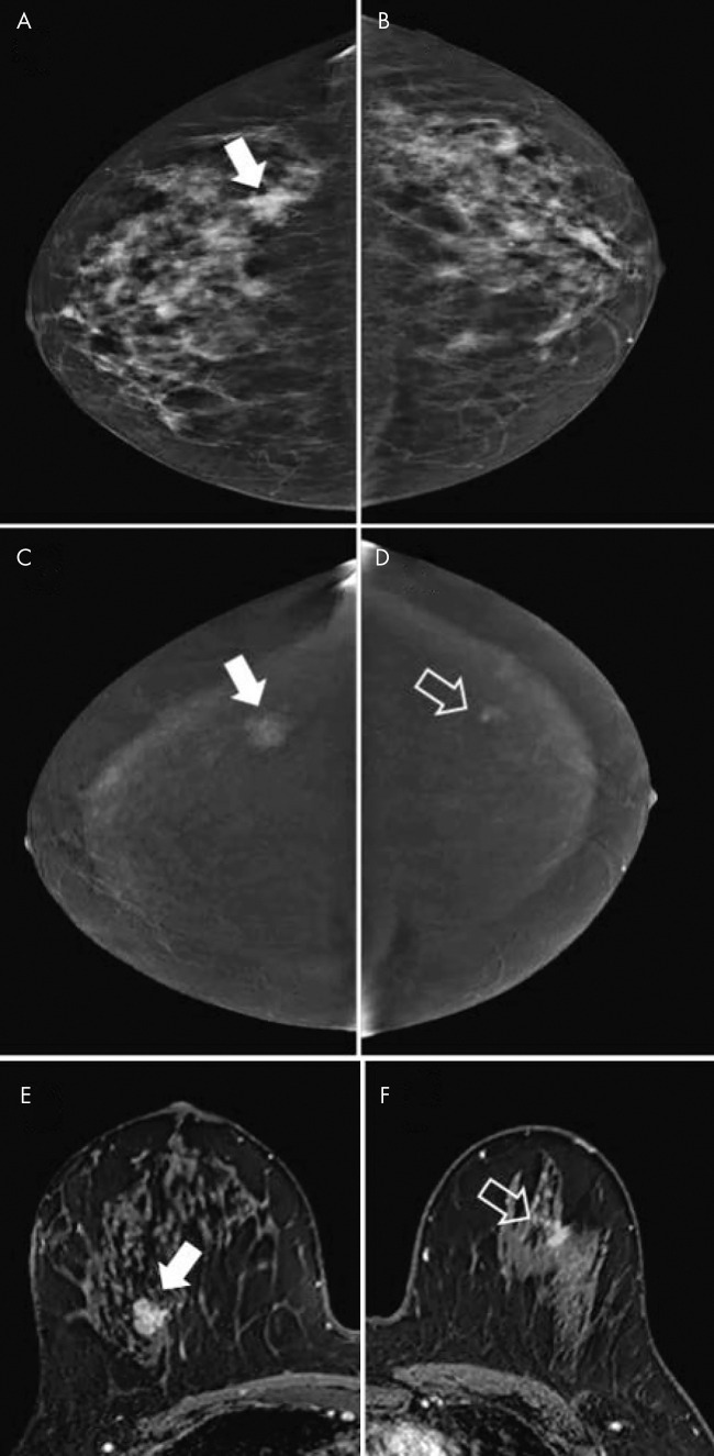
Images obtained for disease extent assessment in 64-year-old woman recalled from screening for irregular mass in right breast. A, Low-energy image of right breast (craniocaudal view) shows irregular mass (arrow). B, No lesion is seen on low-energy image in contralateral breast. C, Image from contrast-enhanced mammography (CEM) (right craniocaudal view) shows enhancement of mass (arrow). D, CEM image shows contralateral irregular enhancing mass (arrow), which was not visible on low-energy image. E, F, Contrast-enhanced MRI scans of, E, right breast and, F, left breast show both lesions (arrow). Lesions were diagnosed as invasive breast cancer of no special type (estrogen receptor positive, progesterone receptor positive, and human epidermal growth factor receptor type 2 negative).

Images obtained for disease extent assessment in 64-year-old woman recalled from screening for irregular mass in right breast. A, Low-energy image of right breast (craniocaudal view) shows irregular mass (arrow). B, No lesion is seen on low-energy image in contralateral breast. C, Image from contrast-enhanced mammography (CEM) (right craniocaudal view) shows enhancement of mass (arrow). D, CEM image shows contralateral irregular enhancing mass (arrow), which was not visible on low-energy image. E, F, Contrast-enhanced MRI scans of, E, right breast and, F, left breast show both lesions (arrow). Lesions were diagnosed as invasive breast cancer of no special type (estrogen receptor positive, progesterone receptor positive, and human epidermal growth factor receptor type 2 negative).