
Figure 1a:
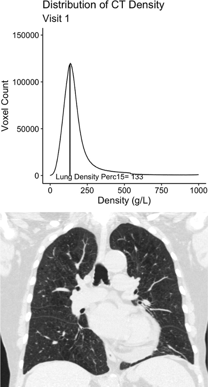
Example 77-year-old female participant from the chronic pulmonary obstructive disease (COPD) gene (COPDGene) cohort with evidence of quantitative emphysema progression but no clear visual progression of emphysema over 5 years who ultimately died during long term follow-up. (a) A representative coronal noncontrast CT image and the associated CT density histogram for the participant at baseline. (b) A representative noncontrast-enhanced coronal CT image and the associated CT density histogram for the participant at the 5-year follow-up visit. Lung density perc15 = volume-adjusted lung density measured at the 15th percentile of the CT lung density histogram.