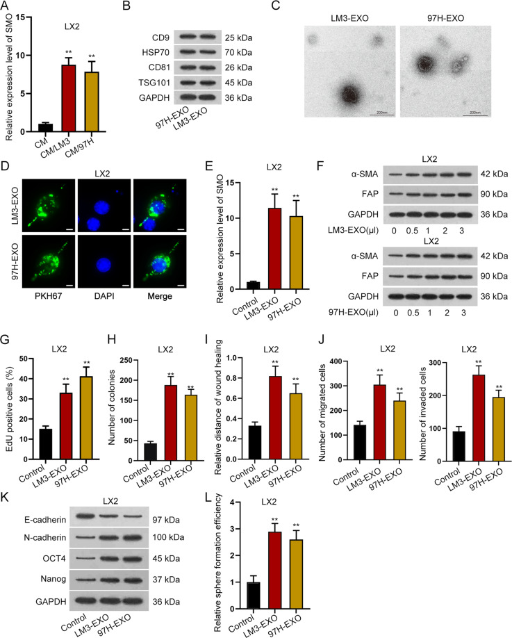
Fig. 2. HCC cells-derived exosomes transmitted SMO and activated HSC.
A qRT-PCR detected the expression of SMO in quiescent HSCs and activated HSCs induced by conditioned medium (CM) of LM3 or 97H cells. B Western blot detected the protein level of CD9, HSP70, CD81, and TSG101 in LM3- or 97H-derived exosomes. C Electron microscope revealed the images of exosomes. D IF staining assay revealed PKH67-labeled exosomes in LX2 cells treated with exosomes from LM3/97H cells (bar value = 15 μm). E qRT-PCR detected the expression of SMO in LX2 cells treated with LM3- or 97H-derived exosomes. F Western blot assay revealed the protein level of α-SMA and FAP in LX2 cells under the treatment of exosomes at different concentrations. G–J EdU, colony formation, wound healing, and transwell assays tested cell proliferation, migration, and invasion in LX2 cells with or without the treatment of LM3 or 97H-derived exosomes. K Western blot analyzed the levels of E-cadherin, N-cadherin, OCT4, and Nanog in quiescent LX2 cells and exosomes-activated LX2 cells. L Sphere formation assay disclosed the effects of LM3- or 97H-derived exosomes on the stemness of LX2 cells. **P < 0.01.

