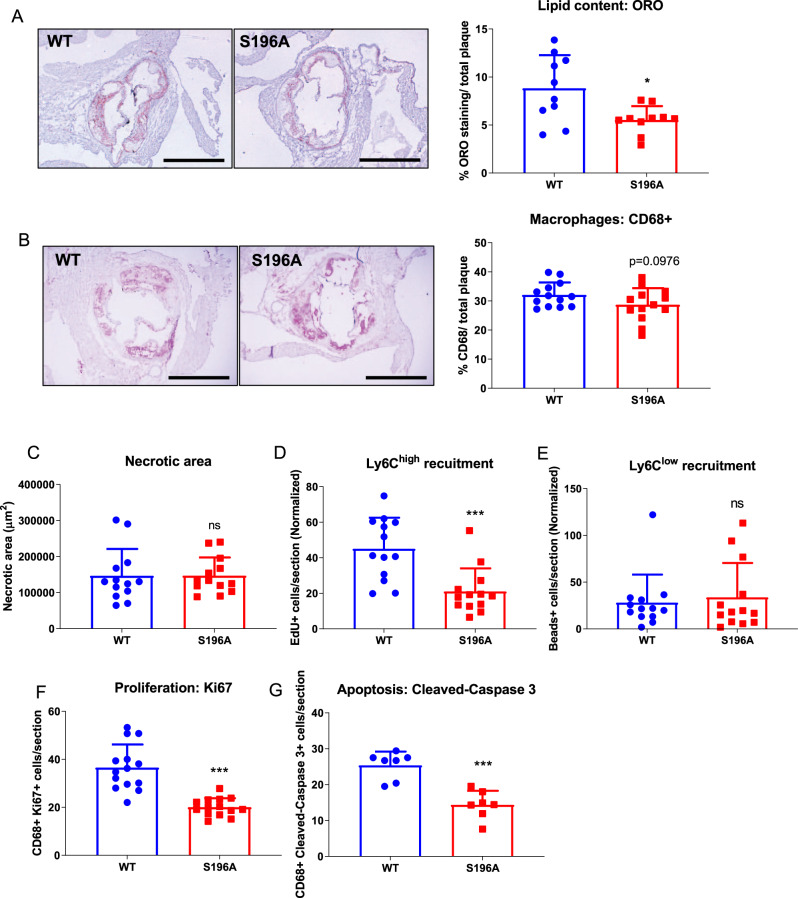
Fig. 2. LXRα S196A decreases lipid accumulation in plaque macrophages, reduces monocyte recruitment and macrophage proliferation, as well as decreases macrophage apoptosis in atherosclerotic plaques.
Representative images and quantification of A Oil Red O, B CD68 immunostaining (scale bars 400 μm), and C necrotic area of aortic root sections from LXRα WT and S196A mice. Histological quantification of monocyte recruitment: D EdU positive Ly6Chigh and E beads positive Ly6Clow cells normalized to the number of positive cells in the blood. F Ki67 (proliferation), and G Cleaved-Caspase 3 (apoptosis) were measured in the CD68+ cells. Data are expressed as mean ± SD (n = 13 except for ORO n = 10 and cleaved-Caspase 3 n = 7) and obtained from independent samples. T test; *P < 0.05, and ***P < 0.001.

