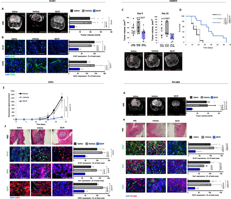
Fig. 7. SELPi treatment delays the growth of murine and human GB tumors in mice.
A Treatment with SELPi reduced tumor growth of GL261 GB tumors in mice. Representative MRI scanning of day 17 post tumor inoculation. Tumor volume was calculated using Radiant software. N = 5 mice per group. Data represent mean ± s.e.m. One-way ANOVA, Holm-Sidak’s method, p < 0.009. B SELPi treatment reduced the proliferation (Ki-67) and microvessel density (CD31) in GL261 GB tumors. Data represent mean ± s.d. Each dot represents the average of three images per mouse. N = 3 mice per group. C Systemic SELPi treatment reduced tumor growth of iAGR53 tumors in mice. Representative images of day 16 scan. P value was calculated by One-way ANOVA test with multiple comparisons adjustment. Tumor volume was calculated using Radiant software. “N” refers to number of mice per group. Center of the box plots shows median values, boxes extent from 25% to the 75% percentile, whiskers show minimum and maximum values. D SELPi treatment prolonged the survival of iAGR53 tumor-bearing mice. N = 15 saline, 8 vehicle, and 12 SELPi (mice per group). P values were determined using two-sided log rank test. E SELPi treatment delayed tumor growth of U251 tumors by SELPi treatment. Data represent mean ± s.e.m. N = 3 saline, 4 vehicle, and 4 SELPi (mice per group). One-way ANOVA, Dunn′s method, p < 0.006. F H&E staining of U251 tumors. Immunostaining demonstrating reduction in proliferation (Ki-67), activated microglia (Iba1), and blood vessels (CD31) in SELPi treated tumors. Data represent mean ± s.d. Each dot represents the average of three images per mouse. N = 3 mice per group. G Tumor volume of PD-GB4 tumors in mice demonstrating delayed growth of PD-GB4 tumors by local SELPi treatment. Representative MRI scanning of day 21 post tumor inoculation. Data represent mean ± s.e.m. Tumor volume was calculated using Radiant software. N = 5 PBS, 3 DMSO, and 5 SELPi. One-way ANOVA, Dunn′s method, p < 0.018. H H&E staining of PD-GB4 tumors. Immunostaining demonstrating reduction in proliferation (Ki-67), activated microglia (Iba1), and blood vessels (CD31) in SELPi treated tumors. Data represent mean ± s.d. N = 3–4 images per mouse, 2 mice per group. Statistical significance of all the immunostaining was determined using one-way ANOVA test with multiple comparisons adjustment. All Scale bars represent 100 µm. Source data are provided as a Source Data file.

