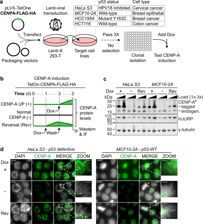
Fig. 1. Inducible and reversible CENP-A overexpression in cells with varied p53 status.
a Scheme for the generation of doxycycline (Dox) inducible CENP-A overexpression cell lines. TetOn-CENPA-FLAG-HA construct was randomly integrated into several indicated target cell lines by lentiviral transfection without selection. After clonal isolation, we tested cells for clear homogenous CENP-A and HA increase by IF in approximately all cells 24 h after the addition of 80 ng/ml of Dox. Cells were also tested to ensure no detectable background HA signal by western or IF, when no Dox was added. b Scheme representing relative CENP-A protein levels over time for TetOn-CENPA-FLAG-HA cell lines. Dox = 10 ng/ml. c Western blot of total cell extracts (TCEs) pertaining to scheme in (b). Primary antibodies indicated on the right. Exogenous CENP-A (tagged) can be distinguished from endogenous CENP-A (endogen.) by its increased molecular weight. # = high sensitivity ECL. y-tubulin used as loading control. See also Supplementary Fig. 1B for western blot analysis corresponding to all cell lines. d Immunofluorescence of paraformaldehyde-fixed cells using anti-CENP-A antibody (green) and DAPI staining (gray). Conditions in parallel with (c). Showing max projection images from a Z-series with zoom on a mitotic cell (DAPI/CENP-A merge) to highlight chromosome arms. Scale bars = 10 µm, Zoom = 2.5 µm.

