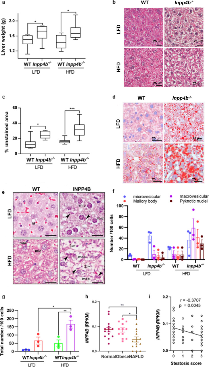
Fig. 4. INPP4B protects mice from liver steatosis.
a Livers were dissected from LFD WT (N = 11), LFD Inpp4b−/− (N = 13), HFD WT (N = 9), and HFD Inpp4b−/− (N = 10) mice. Liver weights in 4 experimental groups (*p = 0.0422 for LFD groups and *p = 0.0374 for HFD groups). b Representative H&E staining of liver sections from designated experimental groups used for steatosis analysis. c Morphometric quantification of the unstained area on H&E stained liver sections (*p = 0.111, ***p = 0.0004). d Representative images of the Oil Red O/hematoxylin staining of frozen liver sections in designated groups. e Characteristic features of hepatocellular steatosis in livers of HFD and LFD Inpp4b−/− males. Examples of microvesicular (mic) and macrovesicular (mac) steatoses, Mallory bodies (closed arrowhead) and pyknotic nuclei (open arrowhead). f Number of individual features per 100 cells. g Total number of steatotic features per 100 cells in each treatment group (*p = 0.0118, **p = 0.0052). h INPP4B expression in Reads Per Kilobase of transcript, per Million mapped reads (RPKM) in normal liver tissue (N = 14), obese (N = 12 patients), and NAFLD (N = 15 patients) in GSE126848 dataset (exact p values are provided in the Source data). One-way ANOVA (Dunnett’s multiple comparison test) was used. i Correlation between INPP4B expression with the steatosis score in patients with NAFLD or NASH. Data were exported from GSE126848.

