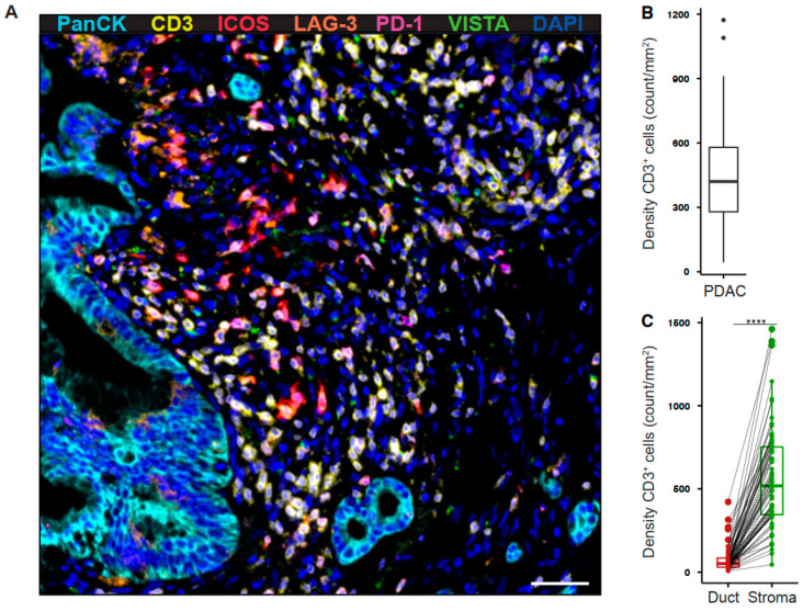
Figure 1.
Tumor-infiltrating T cells reside within the stromal area in pancreatic cancer. (A) Paraffin-embedded human pancreatic ductal adenocarcinoma (PDAC) specimens were stained for PanCK (cyan), CD3 (yellow), ICOS (red), LAG-3 (orange), PD-1 (purple), and VISTA (green). Representative multiplex immunofluorescence image is shown (200×). Scale bar, 50 μm. (B) Quantification of T cell density in whole PDAC and (C) ductal (red) and stromal (green) tissue areas. Each point represents a single patient (total, n = 69). Dot plots and box-and-whiskers (plus min-max), median. Paired Wilcoxon test. p-values ≤ 0.05 were considered significant. ****, p < 0.0001.

