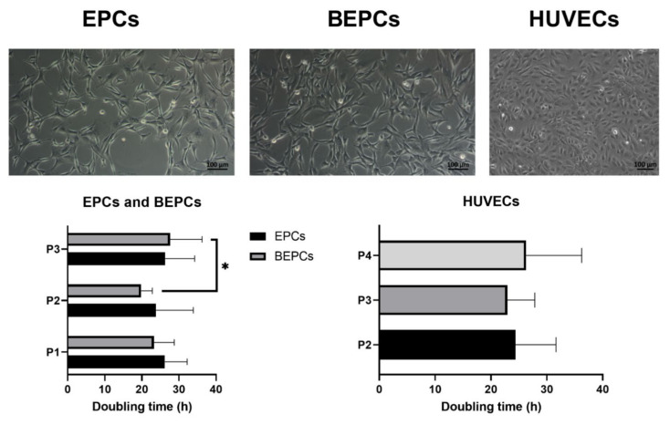
Figure 1.
Cell morphology and population doubling time of the cultured endothelial progenitor cells. Upper part: Rabbit endothelial progenitor cells derived from peripheral blood (endothelial progenitor cells (EPCs)) as well as those derived from bone marrow (BEPCs) showed a spindle-shaped morphology (passage 3), whereas human umbilical vein endothelial cells (HUVECs, passage 4) had a cobblestone appearance as observed under a Zeiss Axio Observer.Z1/7 microscope (magnification at 100×; scale bar = 100 μm). Lower part: Both rabbit (EPCs and BEPCs) as well as HUVEC cell cultures were able to double their concentration after 20–30 h of culture independently of the passage number. The data are expressed as the mean ± standard deviation (SD); *—difference is statistically significant at p < 0.05.

