Figure 2.
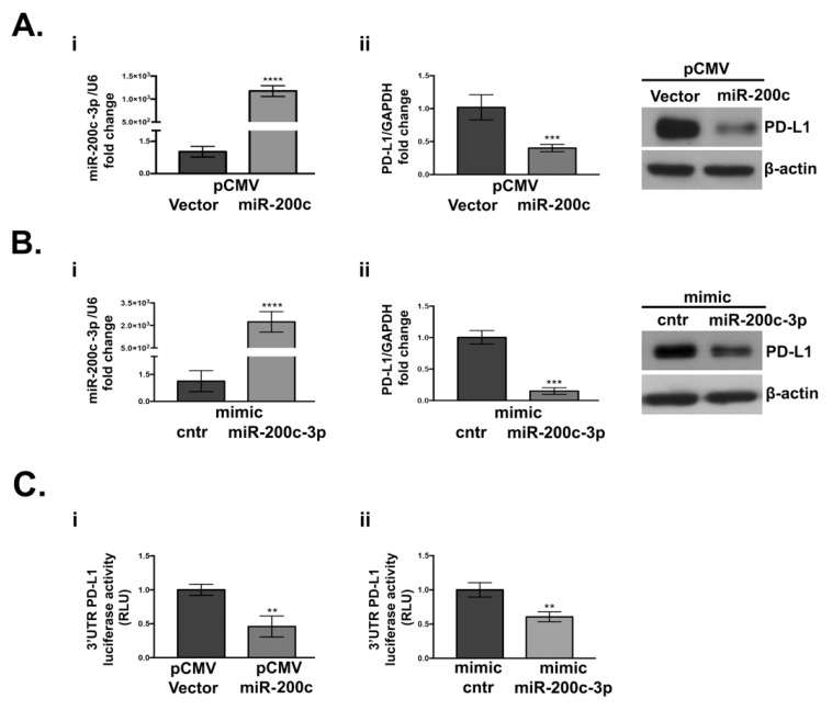
PD-L1 expression decreases upon miR-200c overexpression in SKOV3 cell line. (A) (i): RT-qPCR analysis of miR-200c-3p expression in miR-200c-stably transfected cells (pCMV-miR-200c). (ii): PD-L1 expression is reduced at both transcriptional and protein level, as indicated by RT-qPCR and Western blot (WB). (B) (i): miR-200c-3p expression in transiently transfected cells with mimic miR-200c-3p and mimic control (cntr), at 48 h, by RT-qPCR. (ii): At the same time point (48 h) PD-L1 transcript and PD-L1 protein are downregulated. MiR-200c-3p and PD-L1 fold change expression were normalized to the housekeeping genes RNU6 and GAPDH, respectively. The fold change analysis was performed, and expression values are reported as 2−ΔΔCt. β-actin was used as loading control. (C) (i): Decrease of the 3′UTR of PD-L1 luciferase activity, measured as relative luminometer units (RLU) of firefly-normalized to renilla in miR-200c transfected cells, relative to the empty vector-transfected cells. (ii): A decrease of PD-L1 3′UTR luciferase activity also occurs in the SKOV3 cell line transiently transfected with the mimic miR-200c-3p relatively to the mimic control (cntr). Luminescence detection was performed at 48 h with GloMax Luminometer (Promega). The experiments were repeated at least thrice and in six technical replicates. Two-tailed unpaired t-test was applied in each analysis for statistical significance, ** p < 0.01, *** p < 0.001, **** p < 0.0001.

