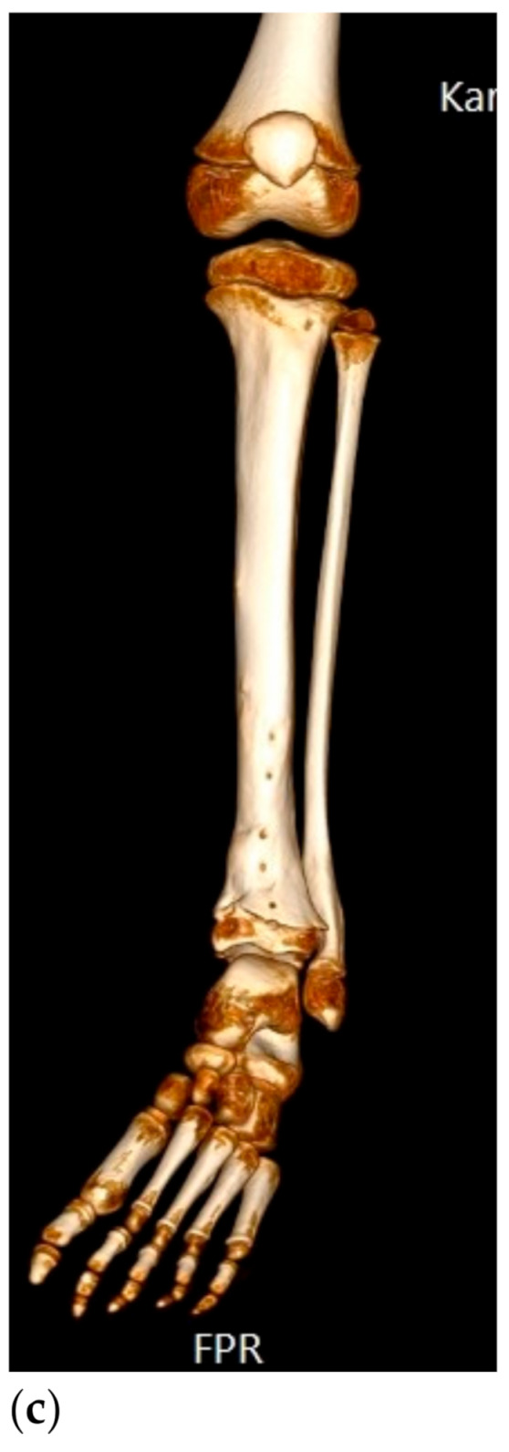
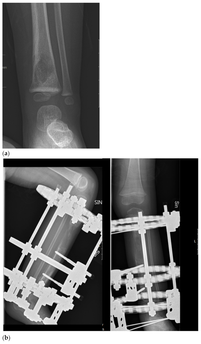
Figure 1.

Biological reconstruction using the bone transport technique (a) A two-year old girl presented to the emergency department unable to walk on her left leg after a mild fall 3 days earlier. She also had a fever and a c-reactive protein count of 87 mg/liter. X-ray showing a lytic lesion located centrally in the distal tibial metaphysis of the left leg. There is a relatively narrow, but indistinct zone of transition. Periosteal reaction involving the medial aspect of the tibia was observed. Open biopsy showed a dense proliferation of small round blue cells in hematoxylin and eosin. Immunohistochemistry showed membranous positivity for the CD99 marker; staining was also positive for S-100 and periodic acid–Schiff (PAS). Fluorescent in situ hybridization (FISH) demonstrated an EWSR1-FLI1 fusion transcript consistent with the diagnosis of Ewing sarcoma. Staging procedures did not show any metastases. (b) Induction chemotherapy with VIDE (vincristine, ifosfamide, doxorubicin, and etoposide) was given according to the Euro-Ewing 2008 protocol. Thereafter, physeal distraction, tumor resection, and segmental bone transport with the use of the Taylor Spatial Frame were carried out. The external fixator was removed after 6 months. (c) Five years after removal of the external fixator, the girl is pain free and is able to run and walk without any limitations. Two deformity procedures have been performed after completing oncologic treatment due to a varus deformity secondary to a physeal arrest in the distal tibial physis. She has no evidence of disease.

