Figure 2.
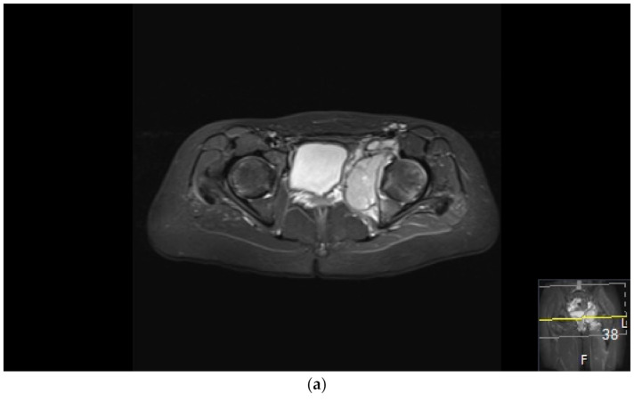
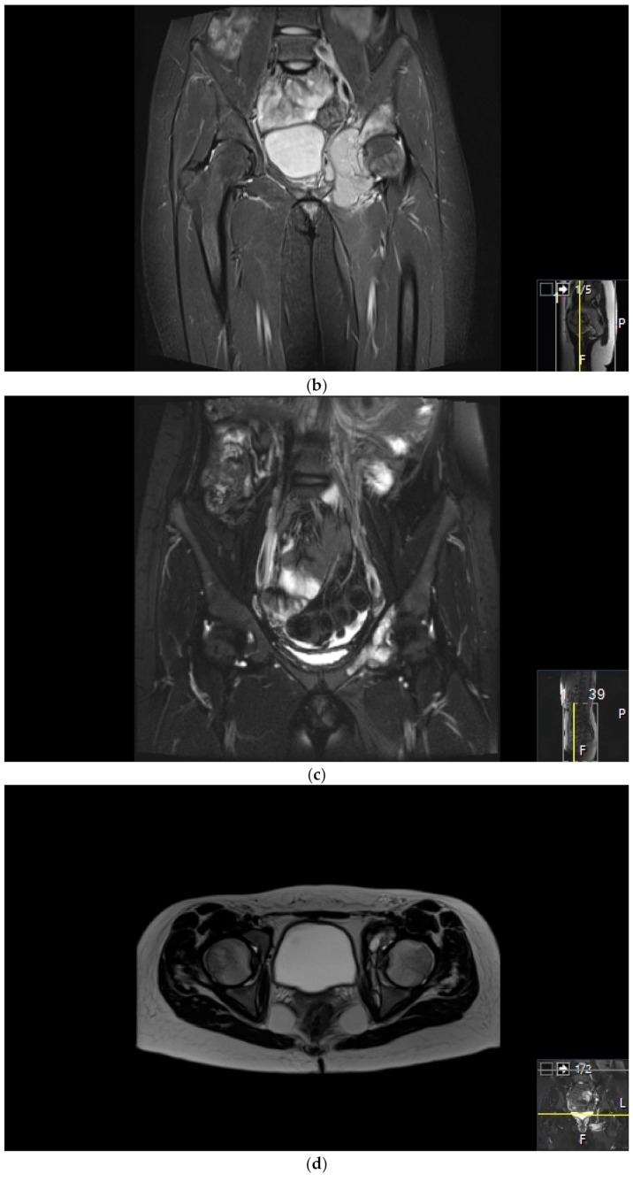
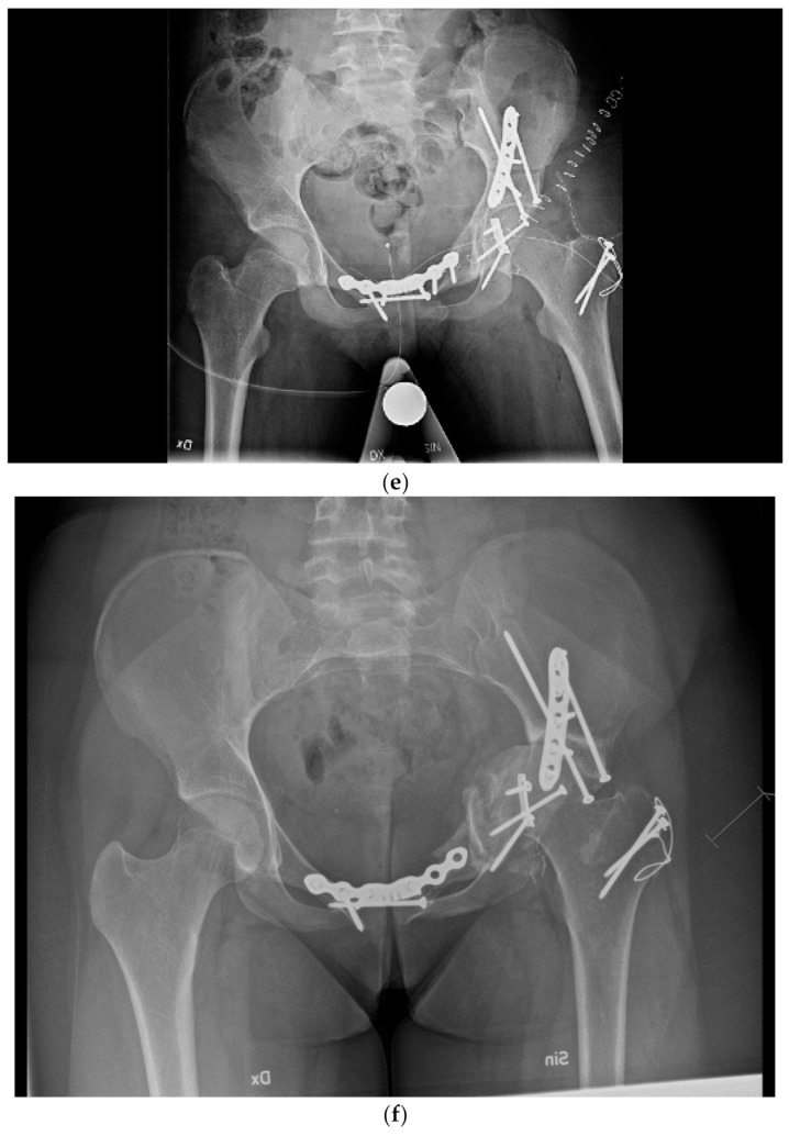
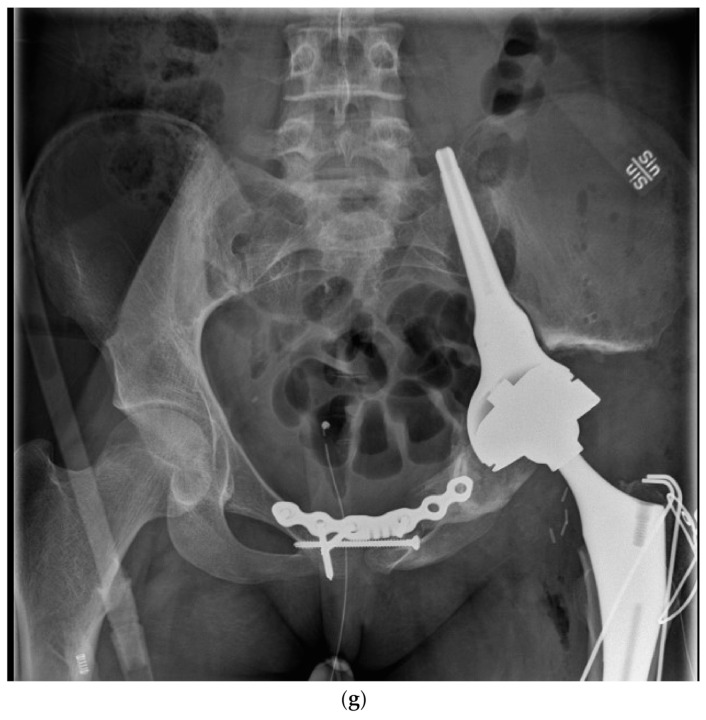
Surgical failure of biological reconstruction after major pelvic surgery, salvaged with endoprosthesis. (a,b) A previously healthy 15-year old girl with 1-year history of left-sided groin pain. MRI of the pelvis (T1 TIRM coronal (a) and axial (b) images) showing a bone tumor involving the left superior ramus of the pubic bone and the periacetabular region of the iliac bone. There is a soft tissue component engaging the obturator internus-externus and adductor muscle. Staging procedures did not show any evidence of metastatic disease. Fine-needle aspiration showed a monotonous small round blue-cell tumor most likely representing Ewing sarcoma. FISH analysis showed an EWSR1-Fli1 fusion transcript confirming the Ewing sarcoma diagnosis. (c,d) After induction chemotherapy with VIDE (vincristine, ifosfamide, doxorubicin, and etoposide), the soft tissue component, as well as the intraosseous extension of the tumor, was significantly reduced, as shown on the coronal (c) and axial (d) T2 TSE FS MRI images. (e) The patient underwent a P2/P3 internal hemipelvectomy, extra-corporeal irradiation with 55 Grey and re-implantation of the autograft. (f,g) One year after primary surgery, the autograft collapsed (f), requiring salvage reconstruction with the Mutars Lumic Cup (g). A year later, the patient is functioning well and remains free of disease.


