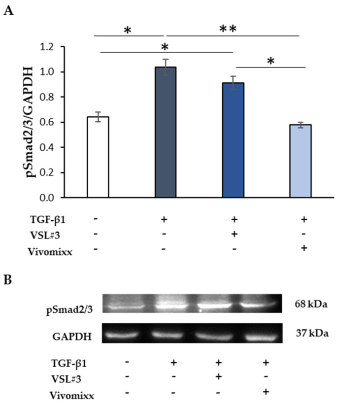
Figure 4.
The effect of the soluble fraction from VSL#3 and Vivomixx lysates on TGF-β1-induced Smad activation. (A) Immunoblotting assay for pSmad2/3 protein expression was performed on CCD-18Co cells incubated for 48 h with TGF-β1 (10 ng/mL) in the presence or absence of VSL#3- or Vivomixx-derived fractions (50 µg/mL). Following densitometric analysis, the obtained values were normalized vs. GAPDH. The data are from two independent experiments in duplicate, and values are expressed as mean ± SEM. For comparative analysis of data, a one-way analysis of variance (ANOVA) with post hoc Bonferroni test was used. * p < 0.05, ** p < 0.01. (B) A Representative image of immunoblotting for pSmad2/3 and GAPDH is shown.

