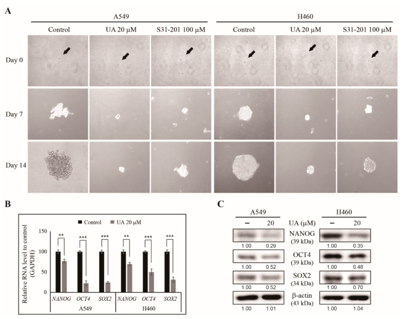
Figure 4.
UA inhibited tumorsphere formation from NSCLC cells. (A) Tumorsphere formation was inhibited by 20 µM UA or 100 µM S3I-201 treatment after the A549 and H460 cells were cultured in DMEM/F12 media containing epidermal growth factor (EGF), basic fibroblast growth factor (bFGF) and B27 for 14 days. Photographs were taken on days 0, 7, and 14. Black arrows indicate the single cells on day 0. (B) RT-qPCR of the mRNA isolated from the A549 and H460 tumorspheres to demonstrate the expression of CSC marker genes after treatment with 20 µM UA for 24 h. The representative expressions of NANOG, OCT4 and SOX2 are shown. The Cp values were normalized to the level of GAPDH mRNA, which was set to 100. (C) Western blotting of the CSC marker proteins, NANOG, OCT4, and SOX2, in A549 and H460 tumorspheres after treatment with 20 µM UA for 24 h. The relative levels of proteins were determined by densitometry and normalized to that of β-actin, which was set to 100. The data were confirmed after repeating the experiment 3 times. ** p < 0.01 and *** p < 0.001 (Student’s t-test).

