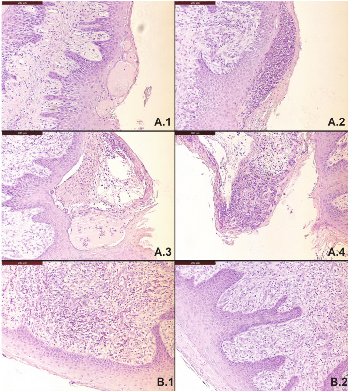
Figure 2.
(A.1–A.4) Histological examination of the rumen wall of cow L1 on parts with a reduced amount of papillae; remaining papillae show formation of exudate filled blisters in the epithelial layer (A.1), and invasion of inflammatory cells (polymorphonuclear leukocytes) into these cavities forming micro-abscesses (A.2,A.3), which leads to ulceration and papillary necrosis (A.4). (B.1,B.2) Histological examination of the rumen wall of cow L6 on local spots with missing rumen papillae and thickening of the rumen wall, and invasion of polymorphonuclear leukocytes (B.1), sometimes combined with hyperkeratosis (B.2).

