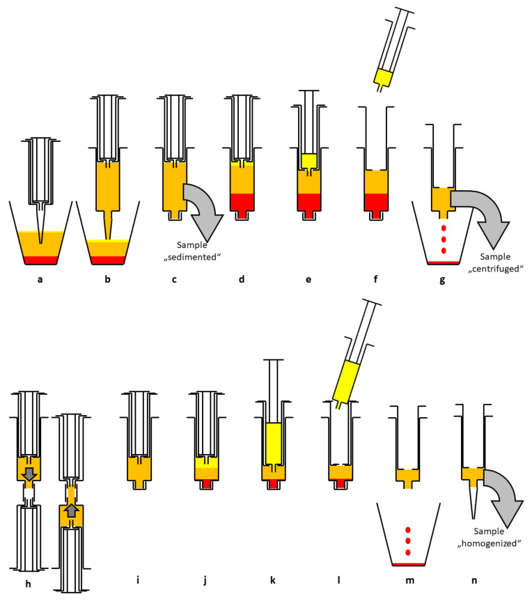
Figure 1.
Schematic diagram of the enrichment process. After sedimentation in the suction container (a) lipoaspirate is transferred to a 15 mL double syringe (b). The sample “sedimented” is taken for analysis (c). After centrifugation (2500 rpm, 4 min) three layers can be seen (d). The upper oil phase is transferred to the small inner syringe (e) and discarded (f). The blood and tumescent solution are discarded as well (g). The sample “centrifuged” is taken for analysis. The syringe is connected to a Tulip-1.4-mm connector and another syringe and the lipoaspirate is emulsified by forcing it through the connector at least 10 times (h). The now homogenized lipoaspirate (i) is centrifuged again at 2500 rpm for 4 min, resulting in three layers (j). The upper oil phase from disrupted adipocytes is transferred to the small inner syringe (k) and discarded (l). The aqueous phase is discarded, too (m). The remaining lipoaspirate contains a high percentage of ASCs and is ready for lipofilling (n). The sample “homogenized” is taken for analysis.

