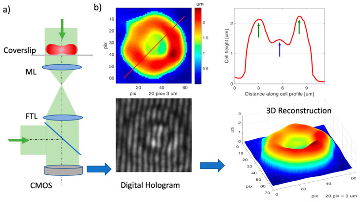
Figure 2.
Digital holographic microscopy (DHM) for 3D RBC imaging. (a) Schematic of the DHM setup. Green arrows: laser beam; ML: microscope lens, TL: tube lens, CMOS: camera sensor; (b) recorded digital hologram on CMOS and 3D reconstruction (bottom); height profile of the cell (top right) along the red line shown in reconstruction (top left).

