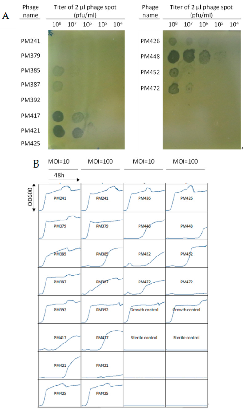
Figure 3.
(A) Plaquing test by the BHI agar overlay method. A total of 2 μL of the ɛ2-phages indicated on the left at 108 pfu/mL were spotted onto a top agar layer containing 108 colony forming units/mL of this patient’s S. epidermidis isolate. (B) “Growth inhibition” curves for the same ɛ2-phages as in (A) with the various bacteriophages to this patient isolates S. epidermidis clinical isolate. Different bacteriophages are indicated by PM241-PM472. MOI refers to multiplicity of infection. Growth control is the clinical isolate with no bacteriophages. Each box has time on the x-axis with end point being 48 h and the optical density (OD) at 600 nm on the y-axis.

