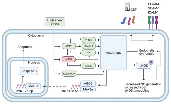
Figure 2.
Autophagy transduces the effects of high-shear stress (HSS) in endothelial cells. HSS promotes autophagy through the activation of AMPK, KLF2, and KLF4, along with the suppression of mTORC1. AMPK in turn directly activates VPS34, Beclin-1, and UKL1, and represses mTORC1. mTORC1 is a repressor of autophagy, which when active binds to and inactivates both ULK1 and ATG13. Autophagy prevents endothelial dysfunction and dampens inflammation by reducing expression of inflammatory mediators (e.g., IL-1, IL-6, GM-CSF) and adhesion molecules (e.g., PECAM-1, ICAM-1, VCAM-1). Moreover, autophagy boosts NO biosynthesis by enhancing phosphorylation of eNOS and blocks eNOS uncoupling and ROS generation. Finally, autophagy mediates anti-apoptotic features by fostering the interaction and nuclear shuttling of Mex3a and miR-126-5p. This allows for the aptamer-like inhibition of caspase-3 function in the nucleus and reduction of apoptosis.

