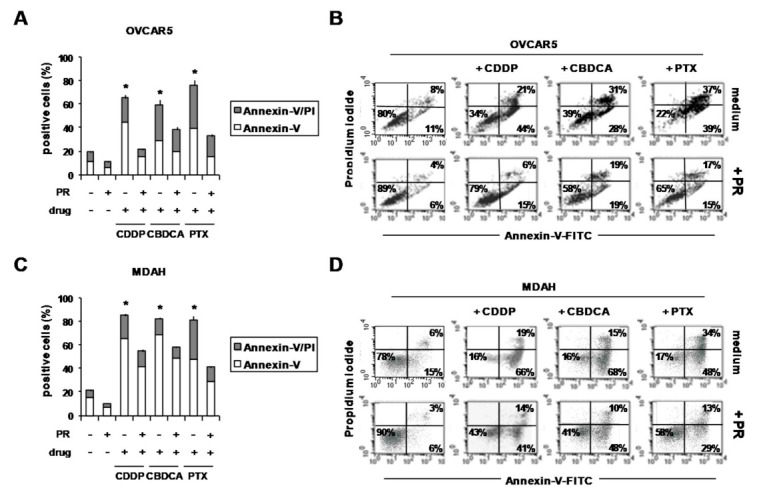
Figure 4.

PR reduced the pro-apoptotic effects of CDDP, CBDCA and PTX in PR-OvCa spheroids. OVCAR5 and MDAH cells were cultured for 5 days with PR (10%) in poly-HEMA-coated wells to form spheroids. Then, cells were treated with CDDP, CBDCA and PTX. After 72 h, OvCa spheroids were dissociated into single-cell suspensions by means of trypsinization and stained with Annexin-V-FITC and PI. (A,C) Histograms showing the percentage of Annexin-V- and Annexin-V/PI-positive cells (B,D) Representative flow cytometric dot-plots showing the percentage of Annexin-V-, Annexin-V/PI- and PI-positive cells. * p < 0.01 (untreated vs. PR-treated cells).
