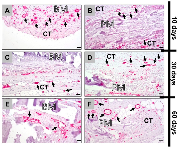
Figure 4.
Immunohistochemically stained slides show detection of CD31-positive endothelial cells into the implantation beds of both bovine (left column: A,C,E) and porcine collagen membranes (right column: B,D,F) at days 10, 30 and 60 after implantation (all images: 400× magnification; scalebars = 20 μm). CT: connective tissue; BM: bovine membrane; PM: porcine membrane; black arrows: CD31-positive vessels.

