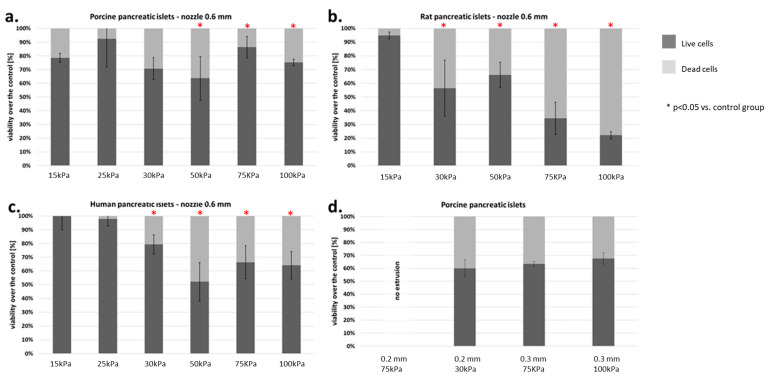
Figure 9.
Viability of islets subjected to the bioprinting process with different pressures and a nozzle with an internal diameter of 0.6 mm. As a carrier material, 3% (w/v) alginate was used, and the vitality of the islets was assessed based on FDA/PI staining and determined in accordance with Equations (5)–(7). Our control consisted of islets right after the isolation. The p-value for each pressure was calculated in comparison to the control group. (a) Porcine pancreatic islets: p-value was determined using Fisher’s method and was 0.22, 0.54, 0.83, 0.042, 0.019, and 0.037 for 15, 25, 30, 50, 75, and 100 kPa groups, respectively. (b) Rat pancreatic islets: p-value was 1.0, 0.001, 0.019, 0.002, and 0.0001 for 15, 30, 50, 75, and 100 kPa groups, respectively. (c) Human pancreatic islets: p-value was 0.95, 0.47, 0.059, 0.019, 0.034, and 0.048 for 15, 25, 30, 50, 75, and 100 kPa groups, respectively. (d) Porcine pancreatic islets: Viability of porcine pancreatic islets subjected to the bioprinting process with different pressures (75 and 200 kPa) and nozzles with an internal diameter 0.2 and 0.3 mm. Statistical significance (p < 0.05) was observed in each experimental group. N = 3. Analyzing the results obtained on the pancreatic islets, we decided to check how the individual cell lines that make up the pancreatic islets behave. We focused our attention on the two largest populations, i.e., α- and β-cells.

