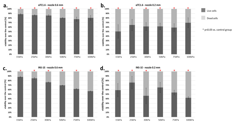
Figure 10.
(a–d) Viability of pancreatic islet cells (α-cells (αTC1.6) and β-cells (INS-1E)) subjected to the bioprinting process with different pressures and nozzles with an internal diameter of 0.6 (Figure 6a,c) and 0.2 mm (Figure 6b,d). As a carrier material, 3% (w/v) alginate was used, and the vitality of the islets was assessed based on FDA/PI staining and determined in accordance with Equation (7). Our control consisted of cells right after the trypsinization process. The p-value for each pressure was calculated in comparison to the control group. In α-cells, p-value was determined using Fisher’s method and statistical significance (p < 0.05) was observed in each pressure for both nozzles. In the case of 0.6 mm diameter nozzles, comparison of the viabilities of control and pressures 15, 25, and 30 kPa showed a loss in cell viability not exceeding 13%. Thus, those pressures can be considered suitable for the bioprinting process. In β-cells, p-value was determined using Fisher’s method, and statistical significance (p < 0.05) was observed in each pressure of both nozzles. In the case of 0.6 mm diameter nozzles, comparison of the viabilities of control and pressures 15, 25, and 30 kPa showed a loss in cell viability not exceeding 18%. Thus, those pressures can be considered suitable for the bioprinting process. n = 3.

