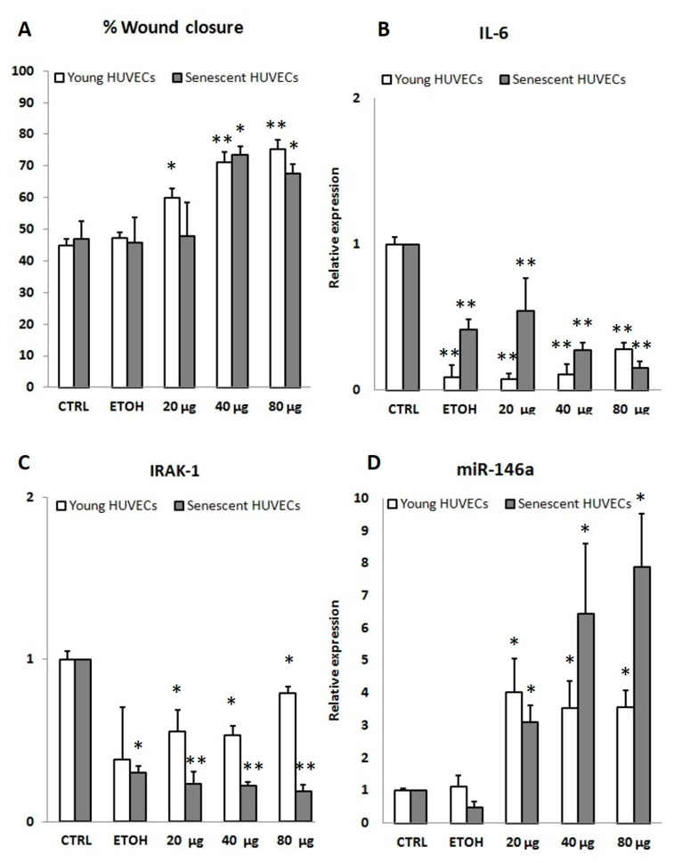
Figure 5.
Wound healing (in % wound closure) in P. spinosa fruit extract (PSF) treated cells and expression levels of inflammation markers assessed in the same cells. Experimental conditions were the same as those employed for anti-inflammaging assays. (A) Percentage of wound healing closure related to CTRL in young (P3) and senescent (P15) HUVECs after 48 h of P. spinosa treatment. Our data showed, both in young and older cells, a significant improvement (up to 70%) of wound healing closure (expressed in percentage related to untreated cells) through cell migration. The same cells were detached and used for the evaluation of expression levels of anti-inflammatory markers, IL-6 (B), IRAK-1 (C), and miR-146a (D). MiR-146a upregulation was observed during PSF extract treatment (20/40/80 µg/mL P. spinosa). PSF extract treatment caused a decreased expression of both IRAK-1 and IL-6, thus revealing a downregulation of the inflammatory response. Results are reported as fold change related to CTRL. Two-tailed paired Student’s t-test: *p < 0.05 vs. CTRL; **p < 0.01 vs. CTRL.

