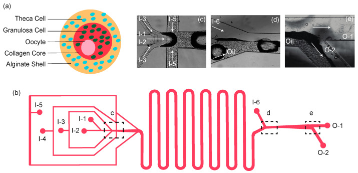
Figure 1.
Design of capsule and device. (a) A schematic illustration of 3D ovarian follicles. The capsule incorporates murine granulosa cells within a soft collagen core surrounded by a separate layer of murine theca cells suspended in a stiffer alginate shell. (b) A schematic view of the microchannel system. Capsules are created at the nozzle where the solutions convene (dotted box, c). Once capsules are generated, they travel through the serpentine channels to allow additional time for gelation and are collected in the top reservoir (O-1). (c) A typical image of the boxed region in panel b showing the flow-focusing areas. (d) A typical image of the boxed region in panel b showing the entrance of the extraction channel. (e) A typical image of the boxed region in panel b showing the exit of the extraction channel. I-1, I-2, I-3, I-4, I-5, and I-6 are the inlets of oocyte, core, shell, Ca-mineral oil emulsion, dispatching, and extraction flows, respectively. O-1 and O-2 are outlets for the aqueous (containing capsules) and oil emulsion flows, respectively.

