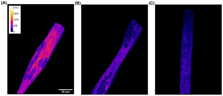
Figure 1.
Qualitative detection with immunofluorescence staining of CB2 receptor in C2C12 skeletal muscle cells. Confocal image of a representative XY acquisition (60× magnification) of (A) differentiated cells incubated with primary CB2 receptor antibody, (B) differentiated cells incubated with CB2 receptor antibody and its blocking peptide (1/1 ratio), and (C) differentiated cells incubated with secondary antibody alone. Scale bar: 36 μm. Secondary antibody, anti-rabbit Alexa 200 Fluor 568, 1:1000. Images are presented in pseudocolor (LUT = fire) to better show the fluorescent intensity variations in the range 0–2552.

