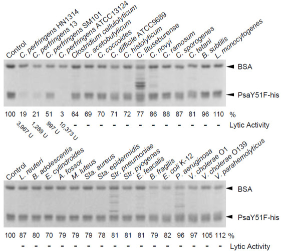
Figure 4.

Cell binding by PsaY51F-his. Cell binding activity was measured as described in the Materials and Methods. Purified PsaY51F-his and the non-binding internal standard, bovine serum albumin (BSA) were combined with cells of a variety of bacterial species (indicated above each gel) or buffer (Control; no cells); the mixtures then were centrifuged, and the resulting supernatants were analyzed by 12.5% SDS-PAGE. Image analysis was used to quantify the intensity (in %; indicated below the gel) of the band of remaining PsaY51F-his following incubation with each bacterium; the intensity of the band in the Control (no cells) was defined as 100%. The lytic activity showed the values in Table 1.
