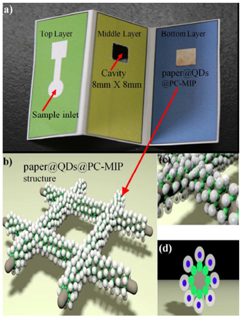
Figure 5.
(a) Photograph of the paper@QDs@PC-MIPs µPADs; (b) Schematic illustration of the structure of the paper@QDs@PC-MIPs component; (c,d) Side and bottom view of enlarged diagram of the structure of the paper@QDs@PC-MIPs on cellulose paper. Green shell: fluorescent quantum dots; white shell: PC imprinting; gray column: cellulose paper. Reproduced with permission from [25]. Copyright 2017 American Chemical Society.

