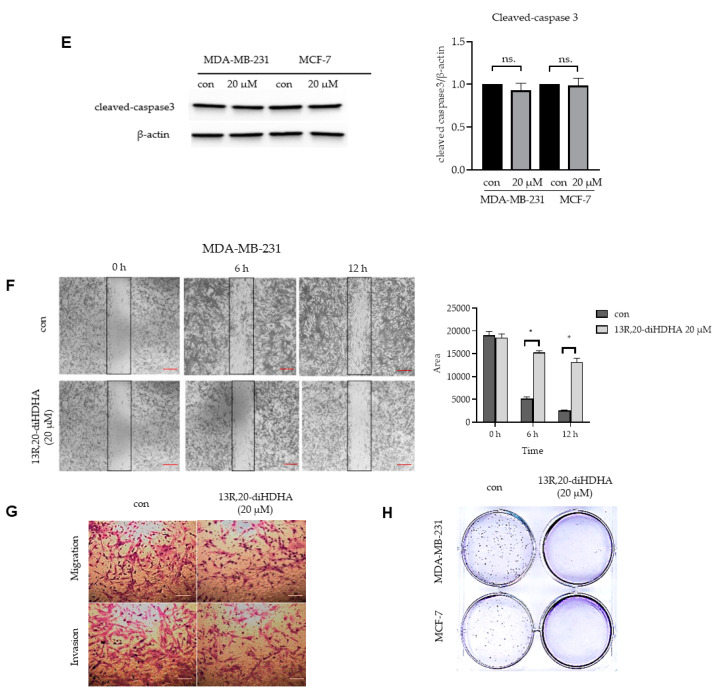
Figure 2.
The effect of 13R,20-diHDHA on mammospheres formation and multiple cancer hallmarks in breast cancer cell lines. (A) MDA-MB-231 and MCF-7 cells were cultured in a 96-well plate with various concentrations of 13R,20-diHDHA for 24 h and cancer cell proliferation was assayed with a CellTiter 96® AQueous One Solution kit. (B) The mammospheres formation efficiency (MFE) was decreased by 13R,20-diHDHA treatment. Mammospheres derived from MDA-MB-231 cells were cultured for 7 days in the presence of 13R,20-diHDHA or DMSO. Image shows the sizes of representative mammospheres, as obtained by microscopy (scale bar: 100 μm). * p < 0.05 versus the DMSO-treated control group. (C) 13R,20-diHDHA prevents mammospheres growth. Mammospheres were treated with 13R,20-diHDHA for 2 days and dissociated to single cells, then equal numbers of cells were plated to fresh dishes. The cells were counted on days 1,2,3 in triplicate and plotted as the mean value. The data from triplicate experiments are presented as the mean ± SD. (D) 13R,20-diHDHA does not induce significant apoptosis of MDA-MB-231 cells. Apoptosis was determined using Annexin V/propidium iodide (PI) staining and FACS. (E) During the mammospheres formation process, 13R,20-diHDHA does not induce a significant change in the level of cleaved caspase 3 (a marker of apoptosis), as determined by western blot analysis. β-actin was used as an internal reference protein. Band density data were used to draw the graph. (F) The migration of MDA-MB-231 cells treated with or without 13R,20-diHDHA (RPMI1640/0.5% FBS) was imaged at 0, 6, and 12 h by a scratch assay (scale bar: 100 μm), and the area was calculated using the Image J software. * p < 0.05 versus the DMSO-treated control group. (G) The cell migration (without Matrigel) and invasion (with Matrigel) of MDA-MB-231 cells exposed to 13R,20-diHDHA were determined by transwell assays (scale bar: 100 μm). (H) Colony formation assays were performed on MDA-MB-231 and MCF-7 cells that had been incubated in 6-well plates and treated with 13R,20-diHDHA (20 μM). Representative colony formation data were collected. The data from triplicate experiments are presented as the mean ± SD.


