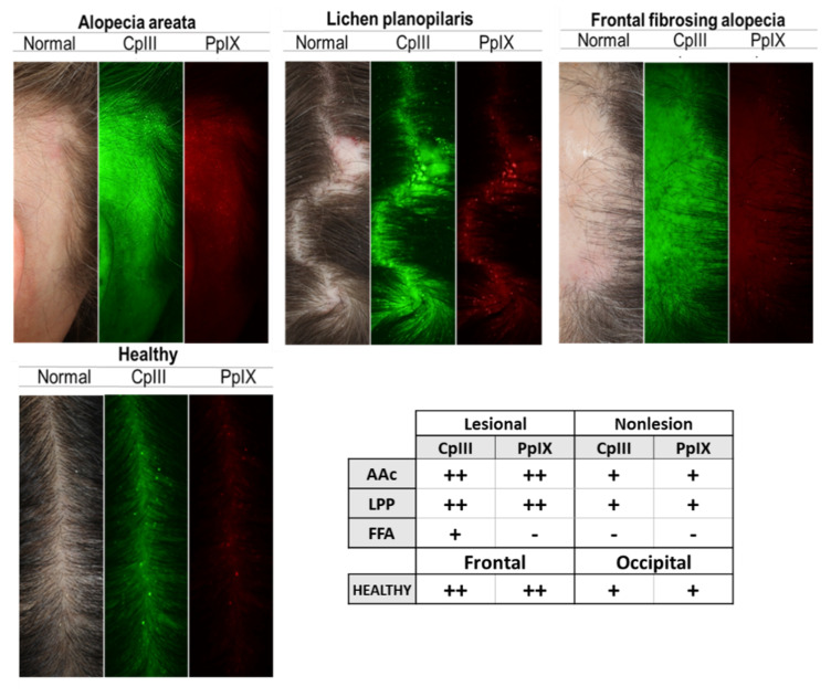
Figure 2.
Cutibacterium acnes presence on the scalp surface of healthy subjects and alopecia patients. Porphyrins, Cutibacterium acnes excretions, fluoresce in UV light, and exhibit circular white spot characteristics. Images from left to right: Standard White Light Image (Clinical Photograph) of the scalp surface (follicular opening); Coproporphyrin (CpIII) Fluorescence Image; and Protoporphyrin (PpIX) Fluorescence Image. Green fluorescence spots correspond to CpIII; red fluorescence spots correspond to PpIX. Table shows the results of the analysis of CpIII and PpIX presence on the scalp, using VISIA CR images. Porphyrins are less numerous on FFA patients. Fluorescence dots × field of view: +, 50–150; ++, 150–250; +++, >250. AAc (n = 7), LPP (n = 6), FFA (n = 6), Healthy (n = 12). LPP, lichen planopilaris; FFA, frontal fibrosing alopecia; AAc, alopecia areata circumscripta.

