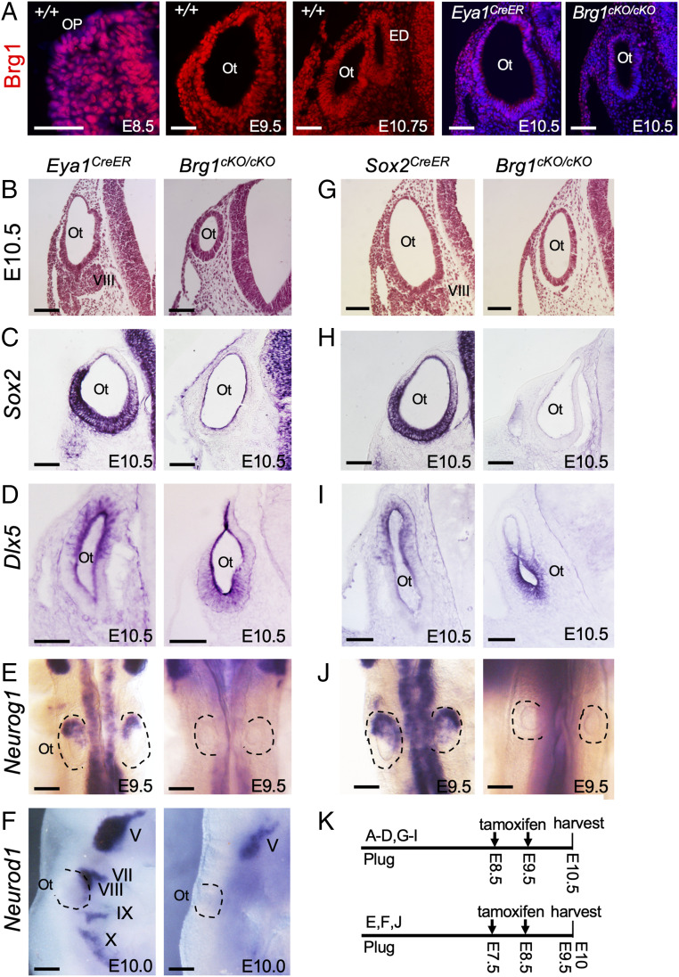
Fig. 1.
Deletion of Brg1 using Eya1CreER or Sox2CreER in otic placode results in lack of neurosensory cell fate specification. Sections are transverse and dorsal is up. For whole-mount images, anterior is up. (A) Anti-Brg1 immunostaining on sections showing Brg1 expression in otic placode (Op) and otocyst (Ot). The Left is a merge of red and Hoechst nuclear staining. (Scale bars, 50 μm.) (B and G) H&E-stained sections of otocyst of Eya1CreER and Brg1cKO/cKO (Eya1CreER;Brg1fl/fl) or Sox2CreER and Brg1cKO/cKO (Sox2CreER;Brg1fl/fl). (Scale bars, 50 μm.) (C, D, H, and I) ISH on sections for Sox2 or Dlx5. (Scale bars, 50 μm.) (E, F, and J) Whole-mount ISH for Neurog1 (dorsal view, E and J) or Neurod1 (lateral view, F) in otic and cranial ganglia. (Scale bars, 120 μm.) (K) Schedules for tamoxifen administration. n = 6 embryos for each genotype. ED, endolymphatic duct; V to X, Vth to Xth cranial ganglion.

