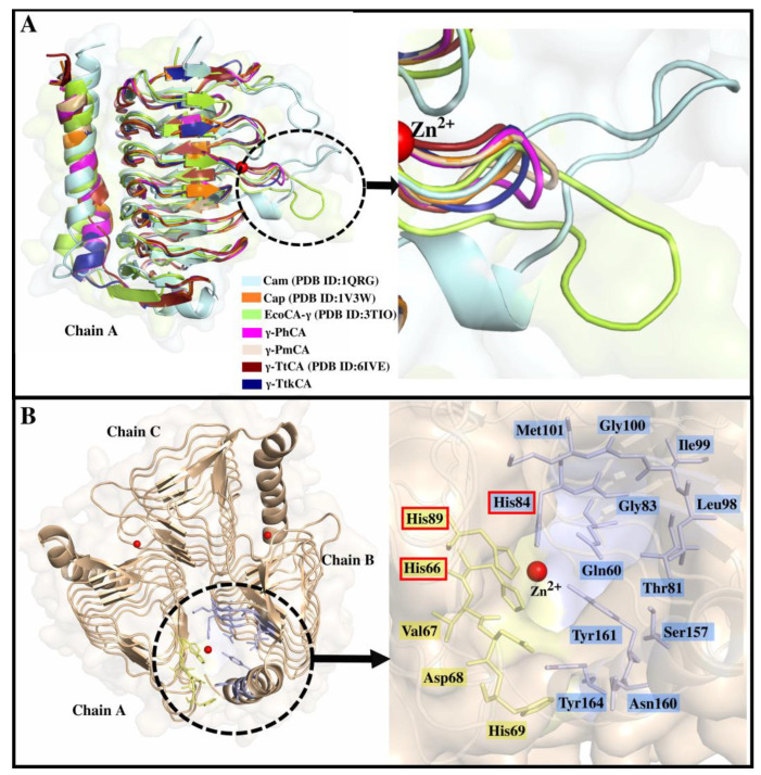
Figure 2.
(A): Aligned monomeric γ-CA structures of Cam (cyan), Cap (orange), EcoCA-γ (green), γ-PhCA (magenta), γ-PmCA (wheat), γ-TtCA (maroon), and γ-TtkCA (dark blue). The loop region indicated in the MSA is shown by the black dotted circle and zoomed into in the image pointed to by the black arrow. (B): Trimeric structure of γ-PmCA with the active site which is located between Chains A and B delimited by the black dotted circle. The black arrow points to the enlarged active site showing the residues that contribute to its formation. Zn2+ is shown as a red sphere and the three His residues coordinating it are boxed in red.

