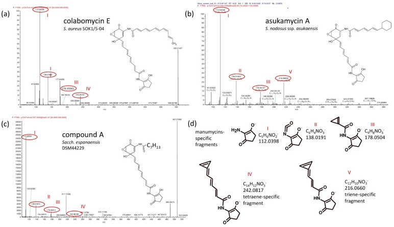
Figure 4.
Comparison of ESI− LC-MS/MS spectra of tetraene colabomycin E (a), triene asukamycin A (b), and compound A (c) The corresponding fragment structures (d) were predicted by Mass Frontier 7.1 software (Thermo Scientific) based on the exact molecular mass. Typical fragments shared by all manumycins containing the C5N unit are shown (I, II, III) together with trienic (V) and tetraenic (IV) lower chain-specific fragments.

