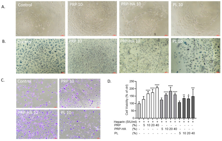
Figure 1.
Effect of platelet-derived products on HUVEC cell behavior. (A). Bright-field optical photography of HUVEC in the presence of heparin, 10% PRP, 10% PRP-HA or PL after 3 days of culture (B). Cellular localization of the MTT formazan in HUVEC incubated for 72 h in different platelet-derived products. HUVEC in control medium and in PL have intracytoplasmic dark granules, while PRP and PRP-HA treated HUVEC show extruded formazan crystals. (C). HUVEC viability assessed by crystal violet staining in control and in PRP-, PRP-HA- and PL-treated cultures. (D). Quantification of the amount of the released dye by absorbance measurement (590 nm). N = 8, * p < 0.05, ** p < 0.01, *** p < 0.005, **** p < 0.001, compared to heparin condition. PRP (platelet-rich plasma), PRP-HA (platelet rich plasma combined with hyaluronic acid), PL (platelet lysates) HUVEC (human umbilical vein endothelial cells). Scale bars: 100 µm.

