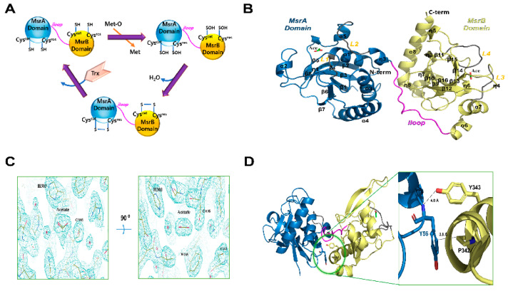
Figure 1.
Crystal structure of HpMsrAB. (A) Cartoon model of the general catalyzed chemical reactions of MsrAB (B) Ribbon diagram of the overall structure of HpMsrABC44S/C318S. The N-terminal domain, MsrA domain (HpMsrA, 34−192) and the C-terminal domain, MsrB domain (HpMsrB, 206−357) are colored sky blue and pale yellow, respectively. The linker region connecting HpMsrA and HpMsrB (residues 193−205), the iloop, is colored magenta. Loops (L1, 42−45 and L2, 181−187) of the HpMsrA domain and loops (L3, 224−229 and L4, 259−266) of the HpMsrB domain are colored gray. Helices (α1–α8), β-strands (β1–β16), and 310-helices (η1–η8) are labeled. Two acetate molecules are represented as stick models colored green. (C) 2Fo-Fc electron density map (1.2 sigma cutoff) around the acetate molecule in the crystal structure. (D) Close-up view of a possible hydrophobic interaction (<4 Å) between Y56 of the HpMsrA domain and P342 of the HpMsrB domain in the crystal structure. The dotted line indicates the distance between the OH of Y343 and the amide N atom of Y56 (~4.0 A), which could not form hydrogen bonds.

