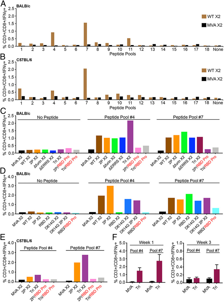
Fig. 4.
CD8+ T cell responses. (A) BALB/c mice and (B) C57BL/6 mice were vaccinated IM with 2 × 10⁷ PFU of unmodified MVA or rMVA expressing WT CoV-2 S at 0 time and again after 3 wk. At 2 wk after the boost, spleen cells were pooled from three to five mice and stimulated with pools of peptides derived from CoV-2 S protein and treated with Brefeldin A. Cells were then stained for cell surface markers with mouse anti-CD3-FITC, anti-CD4-PE, and anti-CD8-PerCP. Cells were subsequently stained intracellularly with mouse anti-IFNγ-APC. CD3+CD8+IFNγ+ cells were enumerated by flow cytometry. (C and D) BALB/c and (E) C57BL/6 mice were primed with the indicated parental MVA or rMVA and boosted with the homologous rMVA or with RBD protein. Splenocytes from four to five mice were combined and stimulated with pool #4 and pool #7 peptides and then analyzed as in A and B. (F) C57BL/6 mice were primed with parental MVA or rMVA Tri. After 1 and 3 wk, the splenocytes of individual mice (n = 4) were analyzed as in A. SDs are shown. X2 refers to splenocytes collected after homologous rMVA boost; RBD Pro in red indicates heterologous boost with RBD protein.

