Fig. 2.
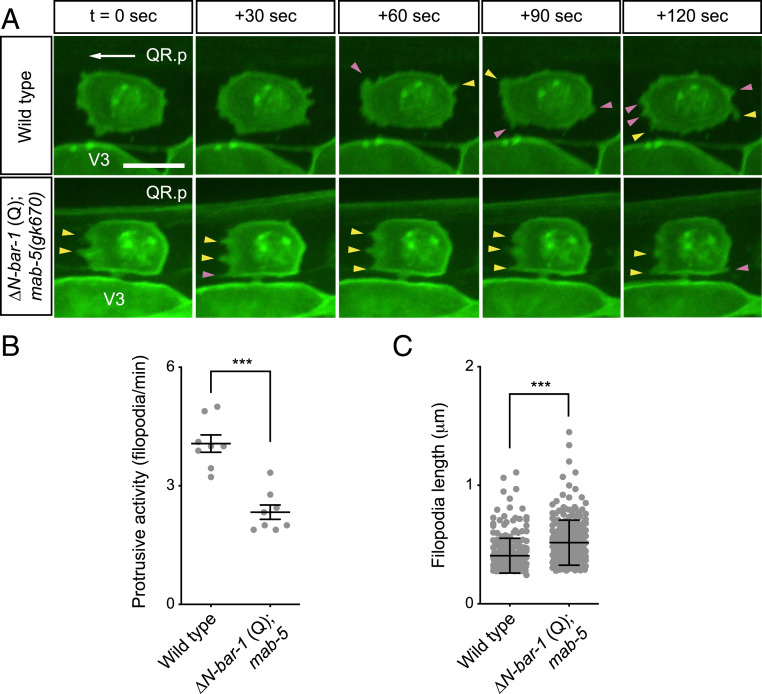
Activation of canonical Wnt signaling influences filopodial dynamics. (A) Time-lapse imaging of QR.p in wild type and ΔN-BAR-1 (Q)–expressing animals. Newly formed filopodia-like protrusions are indicated by pink arrowheads, and stable protrusions are indicated by yellow arrowheads. The seam (V) cells and QR.p are marked with nuclear (H2B) and membrane-localized (PH) GFP (huIs63) (38). The ΔN-BAR-1 (Q)–expressing strain contains mab-5(gk670). Anterior is left and dorsal is up (scale bar, 5 μm). (B) Quantification of protrusive activity, as measured by the number of filopodia-like protrusions formed per minute. Data are represented as mean ± SEM (n = 8 for all genotypes). The statistical significance was calculated using an unpaired t test. (C) Length of filopodia. Data are represented as mean ± SD (n > 245 for all genotypes). The statistical significance was calculated using an unpaired t test. ***P < 0.001.

