Fig. 3.
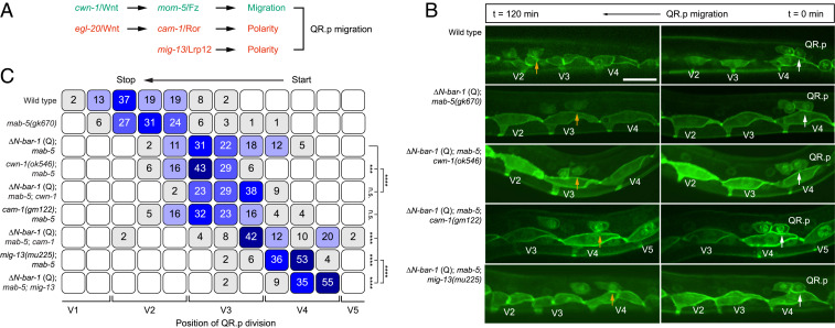
Canonical Wnt signaling counteracts the CWN-1/Wnt–MOM-5/Frizzled–dependent migration pathway. (A) A schematic representation of the parallel Wnt pathways and the mig-13/Lrp12 pathway that control QR.p polarity and migration. (B) Time-lapse imaging of QR.p migration in wild type, ΔN-BAR-1 (Q), and ΔN-BAR-1 (Q) combined with mutations in cwn-1, cam-1, or mig-13. The seam (V) cells and QR.a and QR.p are marked with nuclear (H2B) and membrane-localized (PH) GFP (huIs63) (38). The position of QR.p at time points 0 min and 120 min is indicated by white and yellow arrows, respectively. Anterior is left and dorsal is up (scale bar, 15 μm). (C) Position of QR.p division with respect to the seam cells, indicated as percentiles of the total number of cells analyzed (n ≥ 50 for all genotypes). The ΔN-BAR-1 (Q), cwn-1, cam-1, and mig-13 strains contain mab-5(gk670). The statistical significance was calculated using Fisher’s exact test (n.s., P ≥ 0.05, ***P < 0.001, and ****P < 0.0001).

