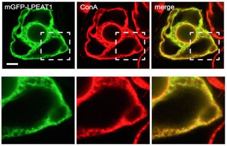
Figure 1.
Subcellular localization of Arabidopsis LPEAT1 in BY-2 cells transiently-transformed via biolistic bombardment with mGFP-LPEAT1, formaldehyde-fixed and processed for confocal laser-scanning microscopy (CLSM), including staining with fluorescent dye-conjugated ConA, serving as an ER marker. Shown are representative images, as well as the corresponding merged image; hatch boxes represent the portion of the cell shown at higher magnification in the panels below. The yellow color in the merged image indicates co-localization between mGFP-LPEAT1 (green) and ConA (red) at the ER. Scale bar, 10 μm.

