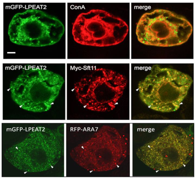
Figure 2.
Subcellular localization of Arabidopsis LPEAT2 in BY-2 cells transiently-(co)transformed via biolistic bombardment with (as indicated by labels) mGFP-LPEAT2 (green) and the Golgi marker Myc-Sft11 (red) or the late endosomal marker RFP-ARA7 (red), or stained with fluorescent dye-conjugated ConA (red), serving as an ER stain. Shown are representative images, as well as the corresponding merged images for each set of (co)transformed cells. The yellow color in the merged images indicates co-localization of two proteins (also shown by arrowheads). Scale bar, 10 μm.

